Models of systems that alternate between two states, such as sleep and awake, are often drawn as consisting of two groups of neurones, each promoting one state, and connected by inhibitory pathways. So the arousal centre inhibits the sleep centre, and vice versa, and when one is dominant the other is silent. The flip-flop switch model suggests that the neurones that cause sleep are in the ventrolateral pre-optic area of the hypothalamus (VLPO) and that they inhibit the arousal system. When the arousal system is active, the VLPO neurones become silent, and the subject is awake (and vice versa for sleep). 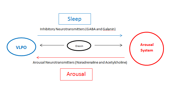
The arousal system is activated from the many sensory systems that cause people to wake up - loud noises, bright lights, somatosensory inputs etc. But in the absence of such inputs there is a basic system controlling the sleep-waking cycle that depends on groups of neurones that alternate in their functions. The ventrolateral preoptic nucleus (VLPO) (analagous to the intermediate nucleus of the human hypothalamus) is active during non-REM sleep, and releases the inhibitory transmitters GABA and galanin on neurons that, when active, promote arousal. The VLPO is in turn inhibited by neurons from brain regions involved in the arousal system. Synchronisation of the EEG is promoted by the VLPO, whereas desynchronisation is promoted by the arousal system (see below). So sleep is an active process - not simply a withdrawal of activity - and the stages of sleep occur in a relatively predictable sequence, and appear to be controlled by different cell groups with different neurotransmitters, driven by the basic flip-flop circuit. The activity of some neurones in the VLPO is promoted by serotonin, and is inhibited by the cholinergic and noradrenergic pathways of the arousal system. Another group of VLPO neurones is inhibited by serotonin. So the roles of these groups of cells and of serotonin has still to be resolved. The arousal system consists of
|
|
| Hypothalamic Nuclei involved in the Sleep/Waking Cycle Top |
|
|
Although not really part of the brainstem reticular formation, several hypothalamic nuclei are involved, along with the brainstem, in the sleep waking cycle. The suprachiasmatic nucleus determines the rhythm of sleeping and waking, and the lateral hypothalamic nuclei that contain orexins have recently been found to be of importance in arousal. Similarly the neurones of the anterior hypothalamus (VLPO) actively promote sleep. Both operate in part by their connections with the brainstem systems. |
|
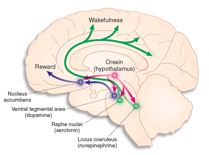
The Orexins (Orexins A and B) are two excitatory neuropeptide transmitters produced from the same gene by groups of neurones in the lateral and posterior hypothalamus. The axons of orexin neurones project to the cerebral cortex and brainstem nuclei, including the locus coeruleus, the raphe nucei and the ventral tegmental area. Orexin neurons excite dopaminergic, noadrenergic, and cholinergic neurones and stimulate food intake, wakefulness and energy metabolism. They are also involved in arousal, and deficiency of orexins can lead to the sleep disorder narcolepsy. |
|
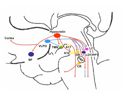
The diagram opposite shows the main interactions of orexin (hypocretin) neurones with cholinergic (BF, PPT), noradrenergic (LC), histaminergic (TMN) and serotoninergic (DR, CR) neurones that promote waking, as well as with the VLPO that promotes sleeping. Orexins seem to act as modulators of the sleep-waking cycle.
|
 * * |